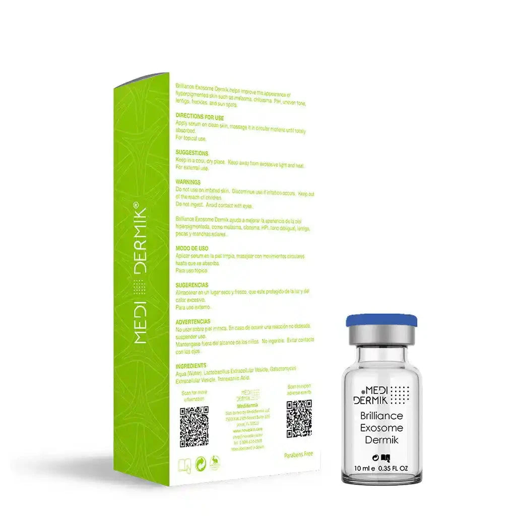
Brilliance Exosome helps improve the appearance of hyperpigmented skin such as melasma, chloasma, PIH, uneven tone, lentigo, fleckless, and sunspots.
It has more than 30 millions of exosomes.
Presentation
5 vials x 10ml (50ml)
Indications
- Melasma.
- Chloasma.
- PIH.
- Uneven tone.
- Lentigo.
- Freckles.
- Sun spots.
How to Use
Brilliance Exosome can be applied by means of:
- Electroporation.
- Iontophoresis (Galvanic Current).
- Dermapen.
- Topical Application Post-Peeling
- Topical Application Post-Microdermabrasion.
- Topical Application with massage.
Ingredients
- Aqua (Water)
- Lactobacillus Extracellular Vesicles 4%.
- Galactomyces Extracellular Vesicles 6%.
- Tranexamic Acid 2%.
Brilliance Exosome Dermik is designed to promote the brightening of the skin with hyperpigmentation issues such as melasma, age spots, lentigo, chloasma, sun spots, and uneven skin tone mediated by the following tranexamic acid mechanisms:
-
Tyrosinase Inhibition:
Reduction of Melanin Production: Tranexamic acid inhibits the activity of the enzyme tyrosinase, which is crucial in melanin production, the pigment responsible for skin color. By reducing this enzyme’s activity, melanin production decreases, helping to lighten dark spots and hyperpigmentation.
-
Plasmin Inhibition:
Reduction of Inflammation: Tranexamic acid acts as a plasmin inhibitor, an enzyme that plays a role in inflammation. Inflammation can stimulate melanin production in the skin, so by reducing plasmin activity, tranexamic acid helps decrease inflammation and, consequently, inflammation-induced hyperpigmentation.
-
Modulation of Growth Factors:
Reduction of Melanocyte Growth Factors: Tranexamic acid can reduce the release of growth factors that stimulate melanocyte activity (the cells that produce melanin).
-
Effect on Keratinocytes:
Blocking Melanin Transfer: In addition to inhibiting melanin production, tranexamic acid can interfere with the transfer of melanin from melanocytes to keratinocytes (skin cells), resulting in less pigment accumulation on the skin’s surface.
-
Synergistic Effect with Other Treatments:
Enhancement of Other Depigmenting Agents: When combined with other depigmenting agents such as Vitamin C 20% Dermik, Glutathione Dermik, niacinamide, or kojic acid, tranexamic acid enhances their effectiveness.
Brilliance Exosome Dermik are tiny, membrane-bound extracellular vesicles secreted by cells that play a crucial role in cell-to-cell communication. Exosomes may contain various bioactive molecules, such as tranexamic acid, proteins, lipids, RNA, and DNA, which can influence cellular processes:
Anti-Aging Effect: Exosomes in general are believed to stimulate collagen production and improve skin elasticity, reducing the appearance of fine lines and wrinkles.
Regeneration: They can promote the repair and regeneration of damaged skin cells, aiding in wound healing and improving skin texture.
Protection against Inflammation: Exosomes may help protect against inflammation, making them useful for conditions like acne and rosacea.
Hydration and Brightening: By enhancing cellular communication, exosomes can contribute to improved skin hydration and a more even skin tone.
When applied to the skin, exosomes can penetrate the outer layers and deliver their cargo to skin cells, thereby modulating cellular functions and promoting skin health.
Exosomes provide a highly effective and versatile delivery system for bioactive compounds, offering advantages in terms of biocompatibility, targeted delivery, protection of cargo, and efficient cellular uptake. These characteristics make them particularly valuable in both therapeutic and cosmetic applications.
When applied to the skin, exosomes can penetrate the outer layers and deliver their cargo to skin cells, thereby modulating cellular functions and promoting skin health.
Exosomes are small extracellular vesicles, typically 30-150 nm in diameter, that are produced by a variety of cell types, including eukaryotic microorganisms such as Galactomyces candidum and Lactobacillus species. These vesicles play a key role in intercellular communication and can carry bioactive compounds such as tranexamic acid, epidermal growth factors, peptides, proteins, lipids, and genetic material. The production of exosomes from Galactomyces candidum or Lactobacillus involves several steps:
Biogenesis: Exosome biogenesis in Galactomyces candidum, as in other cells, occurs through the endosomal pathway:
Endocytosis: The cell membrane invaginates to form early endosomes.
Maturation: Early endosomes mature into late endosomes, also known as multivesicular bodies (MVBs). During this process, inward budding of the endosomal membrane occurs, resulting in the formation of intraluminal vesicles (ILVs) within the MVBs.
Cargo Sorting: Specific bioactive compounds, peptides, proteins, lipids, and nucleic acids are selectively incorporated into these ILVs. The sorting mechanisms involve several protein complexes, including the endosomal sorting complexes required for transport (ESCRT) machinery, as well as ESCRT-independent pathways.
Release: The mature MVBs can follow one of two pathways:
Degradation: Fusion with lysosomes leads to the degradation of their content.
Exocytosis: Fusion with the plasma membrane results in the release of ILVs into the extracellular space as exosomes.
Isolation and Characterization: To study exosomes generated from Galactomyces candidum, they need to be isolated and characterized:
Isolation: Common methods include differential ultracentrifugation, density gradient centrifugation, size exclusion chromatography, and immunoaffinity capture.
Characterization: Techniques such as nanoparticle tracking analysis (NTA), dynamic light scattering (DLS), electron microscopy (EM), and various biochemical assays (e.g., Western blotting for exosome markers) are used to characterize exosomes.
Applications: Exosomes from Galactomyces candidum have potential applications in various fields, including:
Therapeutics: Due to their role in cell communication, they can be harnessed for drug delivery and regenerative medicine.
Diagnostics: Exosomes carry molecular signatures of their parent cells, making them useful biomarkers for disease diagnosis and monitoring.
NovaSkin & MediDermik
Brilliance Exosome